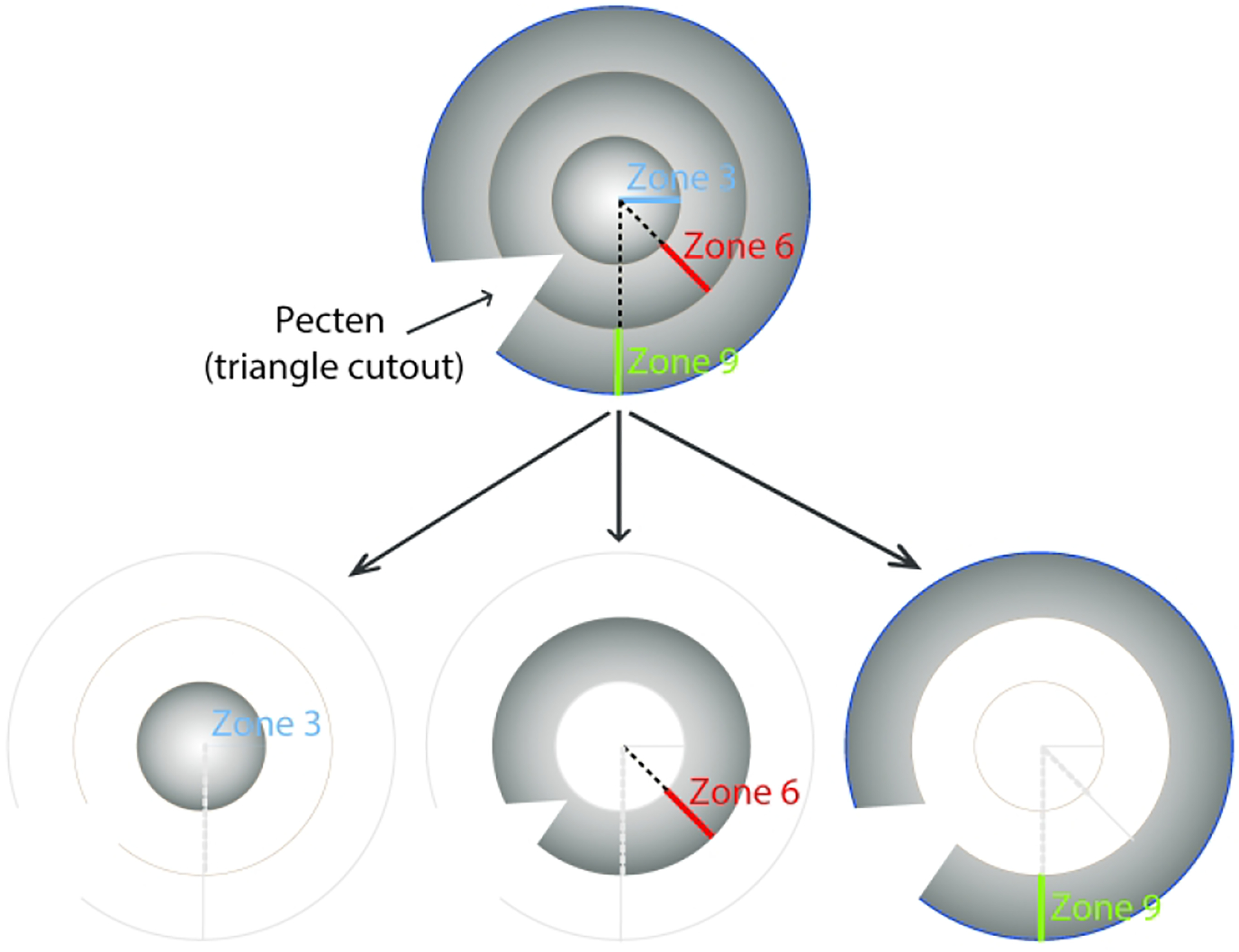
Figure 1.

Diagram showing the origin of the three RPE samples collected from treated and fellow control eyes: a circular 6 mm diameter central zone (zone 3), and two 3-mm wide annular zones, i.e., a midperipheral zone (zone 6), and a peripheral zone (zone 9). The triangle cutout represents the location of the pecten.
