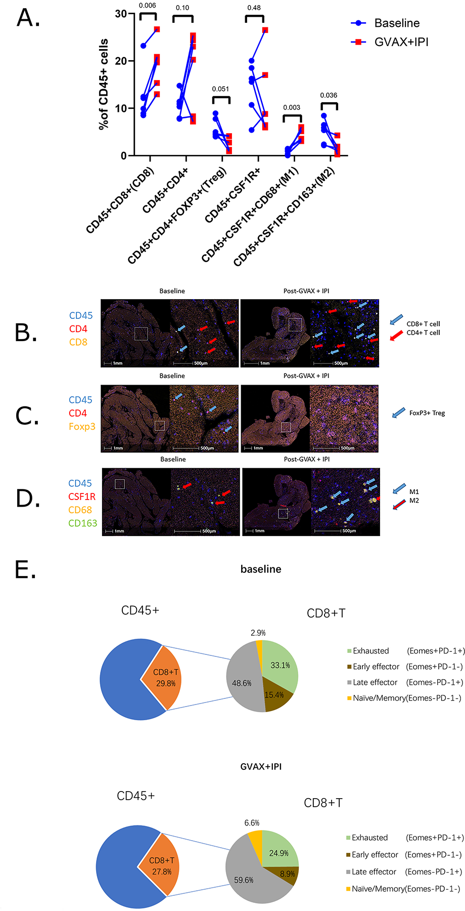
Figure 5. GVAX + IPI promotes an immunostimulatory phenotype in the metastatic pancreatic tumor microenvironment.

A. Longitudinal changes of immune cell composition of CD45+ leukocyte cell densities from baseline (blue circle) to week 7 GVAX + IPI (red square). P values <0.05 indicates statistical significance; Student’s t-tests. B-D. Image visualization by pseudocoloring of baseline and on-treatment FFPE sections of two representative areas in metastatic pancreatic tumor biopsies taken from one Arm A patient who exhibited stable disease. FFPE sections are stained via Multiplex IHC for (B) CD45+CD4+ T helper cells (red arrow), CD45+CD8+ cytotoxic T cells (blue arrow), (C) CD45+CD4+Foxp3+ T regulatory cells (blue arrow), (D) CD45+CSF1R+CD68+CD163+ M2 macrophages (red arrow), and CD45+CSF1R+CD68+CD163− M1 macrophages (blue arrow). E. Comparative analysis of CD8+ T cell functional status at baseline and on-treatment.
