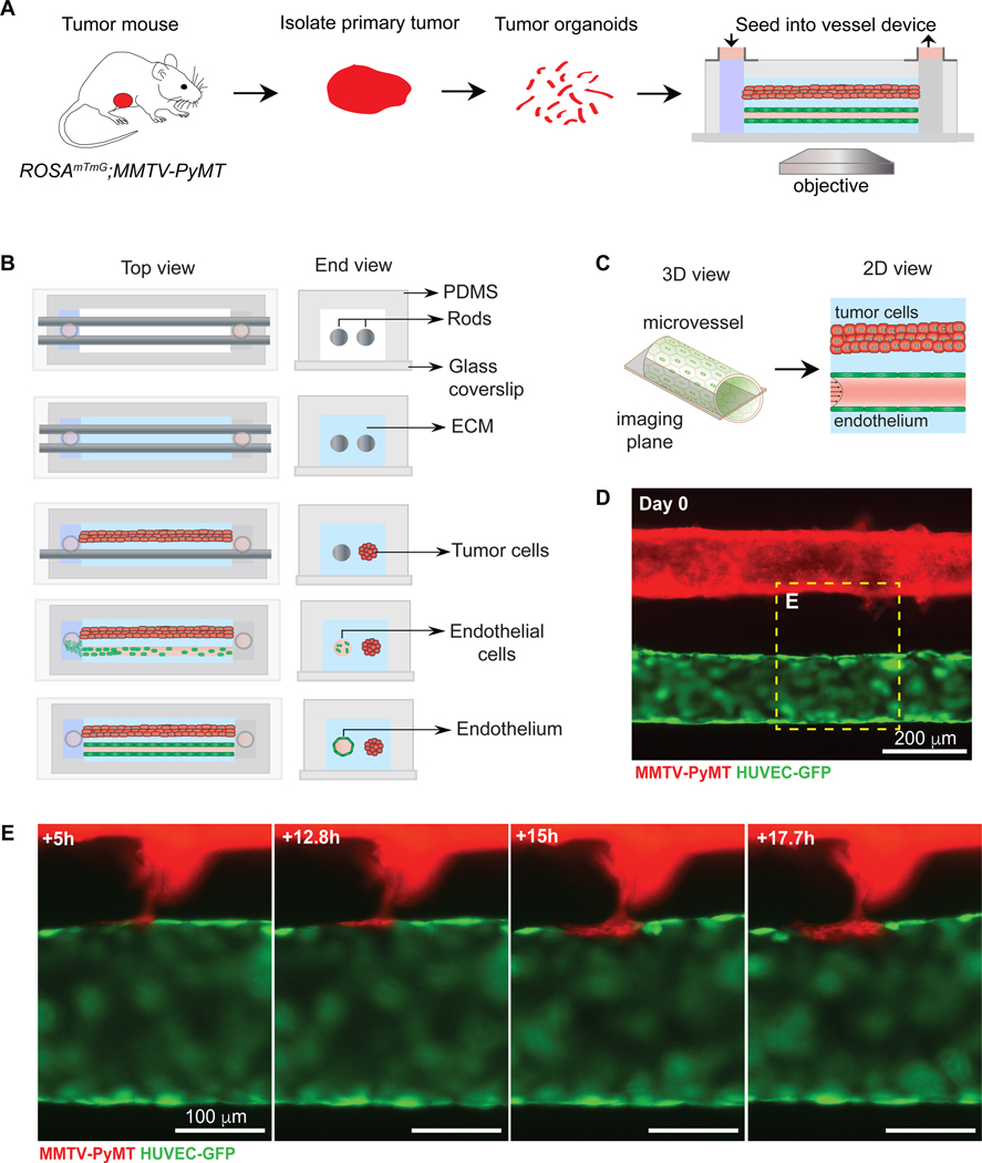
Figure 6. Mosaic vessel formation in a 3D microvessel platform with a parallel tumor-vessel geometry.
(A) Schematic representation of workflow for isolating and imaging organoids in parallel tumor-vessel device. (B) Top- and end-view of microvessel device fabrication. To fix the tumor-vessel distance, a second cylindrical template rod is added into the device, with the collagen solution surrounding both rods. Tumor organoids at a high concentration are injected into one inlet and then the rod is slowly removed, allowing the introduction of the tumor/collagen gel suspension into the cylindrical channel. After the tumor/collagen solution gels, the second rod is removed, resulting in a bare channel which is perfused with media and coated with fibronectin. Endothelial cells are then seeded, resulting in a confluent 3D microvessel. The perfusion system is by gravity flow by differential pressure. Flow is 1 mL/h and shear stress at 4 dynes/cm2. (C) Schematic of the mid-plane of the tumor and vessel. (D) Fluorescence image of a ROSAmTmG; MMTV-PyMT tumor organoid-filled channel and a HUVEC microvessel (GFP), focusing at mid-plane. (E) Frames from a fluorescent time-lapse movie of a collective tumor strand that integrates into the vessel wall, forming a mosaic vessel. The tumor cells are exposed to flow, which is kept at 1mL/hour and shear stress at 4 dynes/cm2. Scale bars: (D) 200 μm, (E) 100 μm.

