
Fig. 3. Newborns have higher representation of Tregs in the lung compared to adults, but their phenotype is similar in the two age groups.
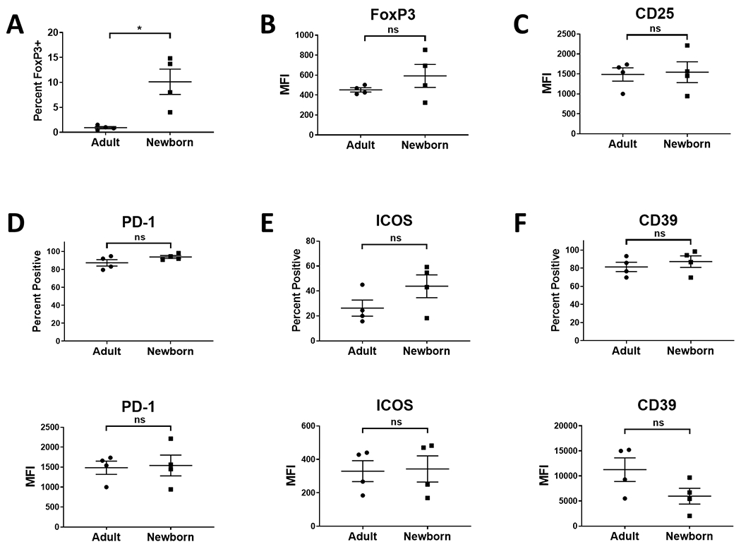
Lung cells isolated from four newborn (4-5 days of age) and four adult (9.6-12.8 years of age) AGM were evaluated for the presence of Tregs. Cells were initially gated on Zombie excluding (live) CD3+ cells. Tregs within this population were identified by co-staining for FoxP3 and CD25. Data for the percentage of CD3+ cells expressing FoxP3 are shown in A. The level of FoxP3 (B) and CD25 (C) on FoxP3+ cells is shown. The percentage of cells expressing the indicated molecule (top panels) and the level expressed by these cells (bottom panel) for PD-1 (D), ICOS (E), and CD39 (F) was also evaluated. Data from four adults is shown in D-F as one animal had a very low percentage of FoxP3+ cells that did not allow confident analysis of subsets. Individuals animals are indicated; the line represents the average±SEM. Significance was determined using a Student’s t-test. **p<0.005. ns=not significant.