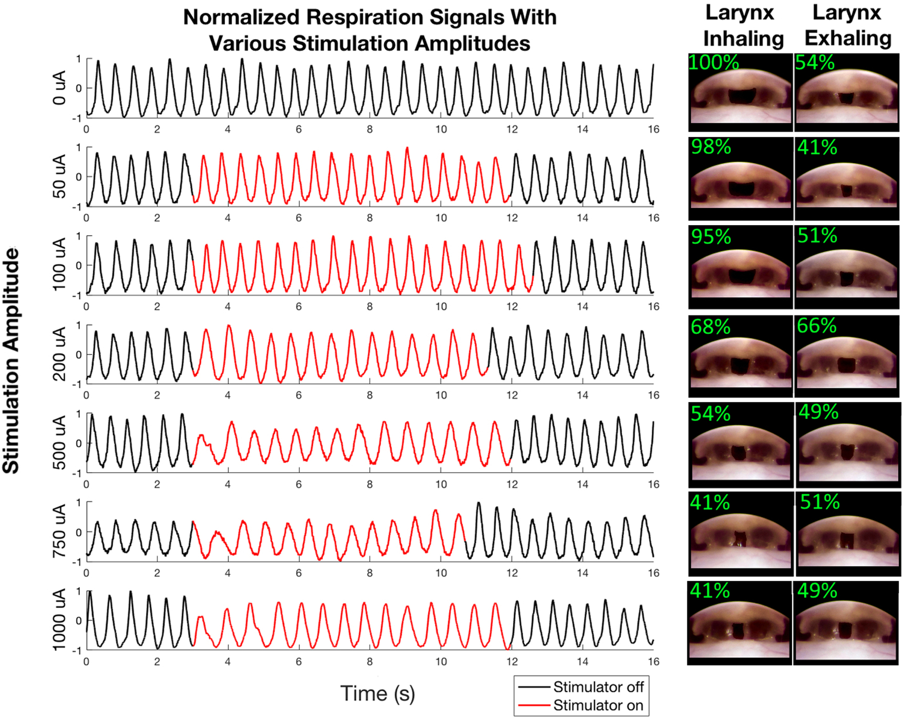
Figure 5:

Effects of laryngeal nerve stimulation. We stimulated both of the recurrent laryngeal nerves with biphasic waveform with the pulse width = 100 μs, pulse repeat time = 500 μs, and variable amplitude. The traces show the normalized respiration signal before, during, and after stimulation. We applied stimulation amplitudes of 0, 50, 100, 200, 500, 750, and 1000 μA as indicated to the left of each respiration trace. The images on the right of the Figure depict the larynx during inhalation and exhalation while stimulating with each amplitude. In each image the region between the vocal folds (i.e. the region in which the trachea is visible past the larynx) has been artificially darkened to make it easier to see the degree to which the larynx is open. The top left of each inset figure shows the extent to which the larynx is open.
