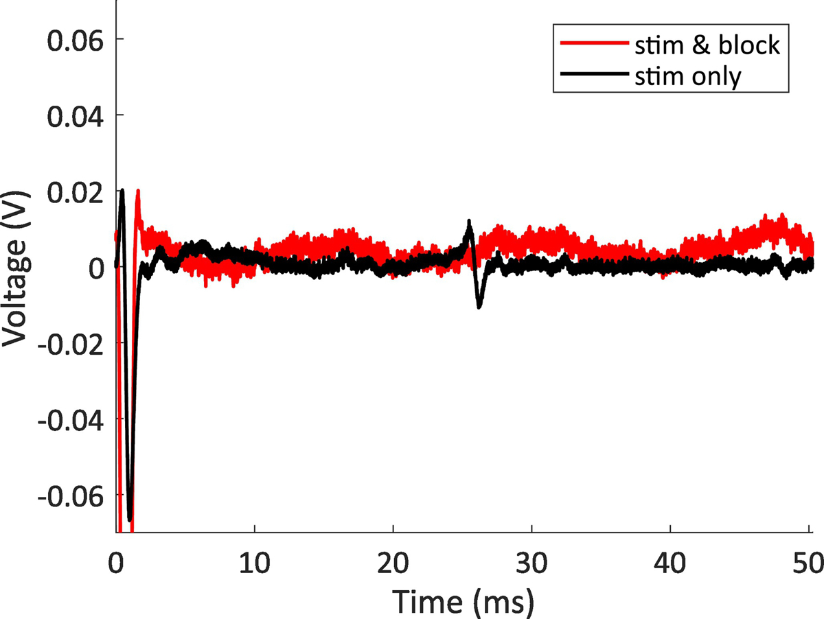
Figure 6:

CNAP blocking example. The data show two stimulation parameters, one with the stimulation waveform applied to the cervical vagus, and one with both the stimulation waveform applied to the cervical vagus and the blocking waveform applied to the gastric branch of the vagus. The nerve response is at approximately 25 ms, so for our interelectrode spacing of 7 cm, the response has a conduction velocity of 2.8 m/s, placing it within the normal range for a B fiber (typically 3.0 m/s or more)(Mirza et al., 2018; Qing et al., 2018; Ward et al., 2015). This sample is representative of the results seen in all animals when only applying blocking stimulation for 5 min or less.
