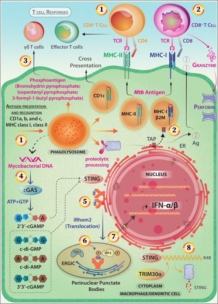
Figure 7.

Antigen presentation, T‐cell responses and activation of the cytosolic surveillance pathway (CSP) in the case of Mycobacterial infection of APCs. (1) Mycobacterial antigens can access both cytosolic and vacuolar antigen‐processing pathways and are presented by the class‐II MHC pathway inducing a potent CD4 response. (2) Presentation in the context of MHC‐I (whereby CD8+ T cell is activated) and CD1 (lipidic antigen) is also reported. (3) Novel phospholigands like bromohydrin pyrophosphate (BrHPP), Mycobacterial antigens (Isopentenyl Pyrophosphate, IPP and non‐prenyl phosphoantigen 3‐formyl‐1‐butyl‐pyrophosphate) are potent elicitors of Vγ9vδ2+ T cells. (4) Mycobacterium activates cytosolic sensor c‐GAS, the STING/TBK1/IRF3 pathway through c‐GAMP and induces Type I IFN‐mediated innate immune responses. (5) Cyclic dinucleotides binding on STING induces its migration from the endoplasmic reticulum (ER) to form perinuclear punctate structures. This intracellular trafficking is mediated by iRhom2. (6) TBK‐1 phosphorylates CTD‐of STING and that results in IRF‐3 recruitment and phosphorylation. (7) The IRF‐3 homodimers translocate to the nucleus to activate the gene transcription of type‐I IFNs. (8) TRIM30α, which is a negative‐feedback regulator of STING via K48‐linked polyubiquitination, marks it for proteasomal degradation. (9) Additionally, Mycobacterium actively employs SecA2 and ESX‐1 secretion systems for releasing RNA into host cells and elicits IFN‐β production through STING and IRF3 activation. Mycobacterial RNA activates the RIG‐1‐MAVS‐TBK1‐IRF‐7 pathway (not shown).
