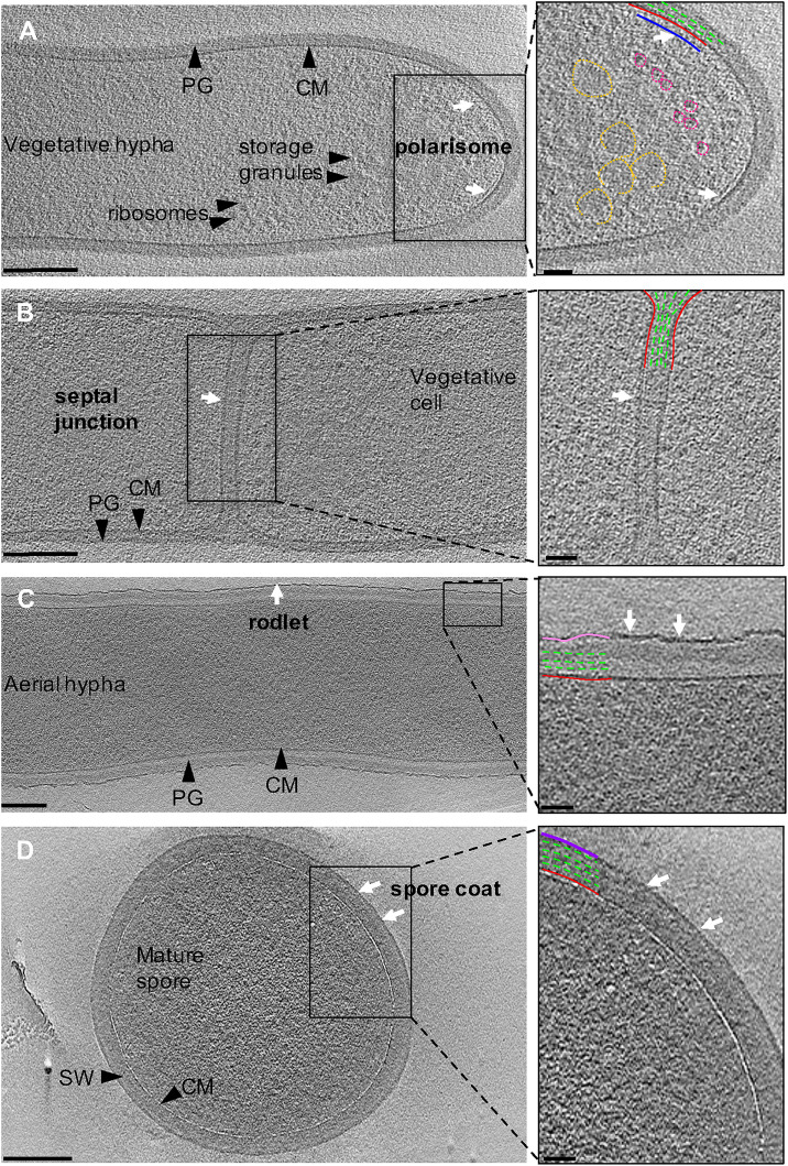
FIGURE 3.
Ultrastructure of Streptomyces. Tomographic slices through S. albus cells at different stages of sporulation. (A) Vegetative hypha. The cytoplasmic membrane and peptidoglycan are shown in red and green, respectively. Putative features include: polarisome (blue, white arrows), glycogen storage granules (yellow), and ribosomes (pink). (B) Vegetative septum between two neighboring cells. A putative septal junction is highlighted with white arrows. (C) Aerial hypha reveal the expected rodlet layer on the surface (pink, white arrows). (D) Mature spore surrounded by a 10-nm thick layer, supposed to be a spore coat (purple, white arrows) on the surface of the spore wall (SW). Insets show a magnified image of the boxed areas. Scale bar = 200 nm, inset 50 nm.

