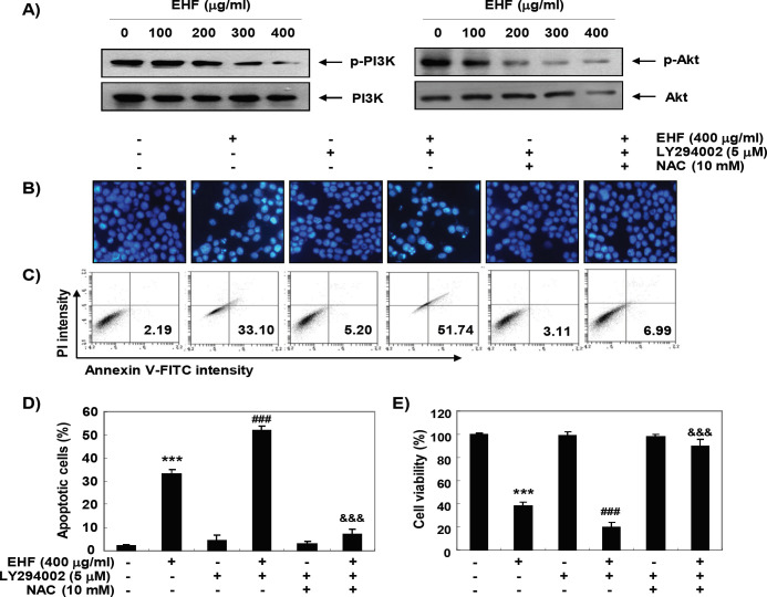
Figure 5.
ROS-Dependent Inactivation of PI3K/Akt Signaling Pathway by EHF in B16F10 Cells. (A) After 48 h treatment with the indicated concentrations of EHF, the cell lysates were prepared and the expression of PI3K and Akt proteins was evaluated by Western blot analysis with whole cell lysates. (B-E) The cells were pre-treated with 10 μM LY294002 and/or 10 mM NAC for 1 h and then treated with 400 μg/ml EHF for further 48 h. (B) The DAPI-stained nuclei were then observed with a fluorescence microscope. The results shown are representative of three independent experiments. (C) The cells were collected and stained with FITC-conjugated Annexin V and PI for flow cytometry analysis. (E) The percentage of apoptotic cells are shown as the mean ± SD (n=3). The statistical analyses were conducted using analysis of variance between groups (***P<0.0001 compared to control; ###P < 0.0001 when compared to EHF-treated cells; $$$P < 0.0001 when compared to EHF and LY294002-treated cells). (E) Cell viability was measured by MTT assay. Each bar represents the mean ± SD of three independent experiments (***P<0.0001 compared to control; ###P < 0.0001 when compared to EHF-treated cells; $$$P < 0.0001 when compared to EHF and LY294002-treated cells).

