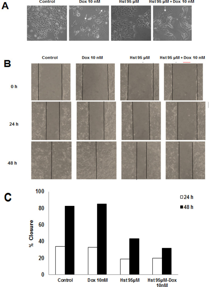
Figure 4.
Effect of Hst, Dox, and Their Combination on Lamellipodia Formation and Cells Migration. An inverted microscope was used to observe lamellipodia formation after treatment with 100x magnification. (A), Morphological change after Hst, Dox, and the combination treatment. White arrow showed lamellipodia of a cell, and observation was done at a certain interval of time; (B), Inhibition of cell migration induced by Dox 10 nM was examined using scratch wound healing assay. Scratch area was observed at a certain time using an inverted microscope at 100x magnification; (C), Percent closure after Hst, Dox, and the combination treatment at 24 and 48 h measured using ImageJ software

