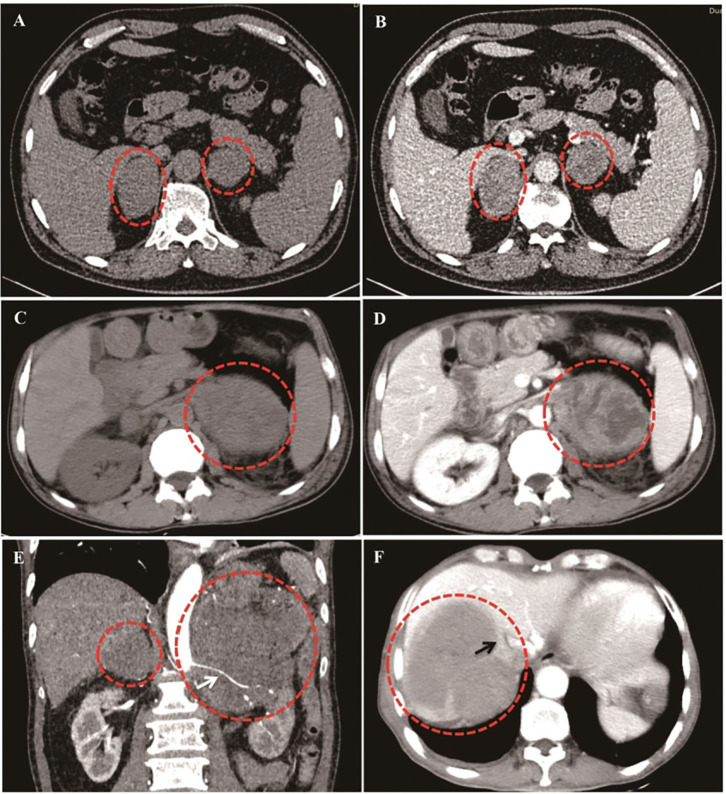
Figure 2.
Different CT imaging features of PAL patients. (A,B): Bilateral PAL with oval/round shape, well-defined edge, moderately enhanced in parenchyma phase, and homogeneous enhancement patterns. (C,D): Bilateral PAL with liquidation/necrosis in the left tumor mass, which was obvious after contrast by iodamide. (E): Lymphoma cells surrounded the renal artery without invasion (White arrow). (F): Right lymphoma mass invaded inferior vena cava (Black arrow).

