. 2020 Sep 9;16(4):562–572. doi: 10.3988/jcn.2020.16.4.562

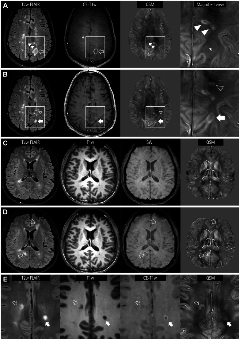
Fig. 2. Longitudinal appearance of QSM rims in MS lesions. A and B: A 44-year-old male patient shows paramagnetic rims (hollow arrow) or nodular enhancements in hyperintense FLAIR lesions (A). A lesion showing a contrast-enhanced rim is slightly paramagnetic (asterisk in the magnified view), suggesting the loss of the diamagnetic myelin tissue. Note the rim or solid paramagnetism in the subcortical FLAIR lesions (solid arrowheads). In follow-up MRI performed 5 months later (B), the size of the FLAIR lesion has decreased (solid arrow) and the enhancement has cleared. The QSM image shows a distinct paramagnetic rim. Additionally, a new periventricular white-matter FLAIR lesion (hollow arrowhead) with a paramagnetic rim is evident. C and D: Another pattern of a new QSM rim lesion in a 23-year-old male MS patient. In the first MRI (C), a small left frontal periventricular lesion is paramagnetic, but it is not accompanied by a paramagnetic rim. At a 9-month follow-up (D), two FLAIR lesions are newly seen (hollow arrows) with paramagnetic rims, with matching dark signals in SWI. E: Another 23-year-old female MS patient shows two subcortical FLAIR lesions with paramagnetic rims in QSM. One lesion shows a complete paramagnetic rim, which is well matched with the enhancement (solid arrow). The other lesion with an irregular and incomplete QSM rim does not exhibit enhancement (hollow arrow). FLAIR: fluid attenuation inversion recovery, MRI: magnetic resonance imaging, MS: multiple sclerosis, QSM: quantitative susceptibility mapping, SWI: susceptibility-weighted images.

Fig. 2