. 2020 Sep 9;16(4):562–572. doi: 10.3988/jcn.2020.16.4.562

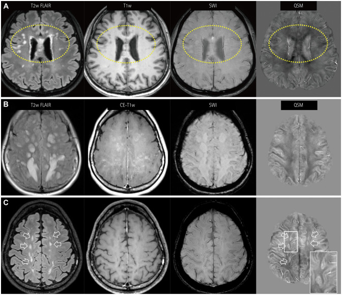
Fig. 3. QSM findings for NMOSD lesions. A: NMOSD lesions without paramagnetic rims in QSM, showing many nodular periventricular and subcortical white-matter lesions in both frontal areas (dotted circles). These lesions do not have dark signals in SWI nor susceptibility changes. B and C: Atypical paramagnetic rims of NMOSD lesions are noted in one patient. The first MRI session (B) shows confluent hyperintense FLAIR lesions. The lesions show patchy or geographic enhancement without susceptibility changes. Follow-up MRI performed 1 year later (C) shows regressed FLAIR lesions (hollow arrows) and enhancement. Some of these lesions show paramagnetic rims in QSM. Note the similarity of the paramagnetic rims (magnified view, box in QSM) with the multiple sclerosis lesions. FLAIR: fluid attenuation inversion recovery, MRI: magnetic resonance imaging, NMOSD: neuromyelitis optica spectrum disorder, QSM: quantitative susceptibility mapping, SWI: susceptibility-weighted images.

Fig. 3