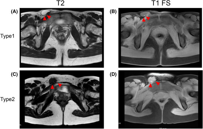
FIGURE 2.

Two types of inguinal endometriosis revealed by magnetic resonance imaging. Red arrowheads denote inguinal endometriosis. A, T2‐weighted axial image shows cystic lesions in the right groin. B, Fat‐saturated T1‐weighted axial image shows the hyperintense nodule in the wall of the cystic lesions. In this case, endometriosis exists and endometriotic lesion exists at the wall of a hernia sac or hydrocele of Nuck’s canal. C, T2‐weighted axial image shows the right inguinal mass (isointense with muscle). D, Fat‐saturated T1‐weighted image shows hyperintensity in the nodule. In this case, endometriotic lesions exist in the solid fibrotic mass
