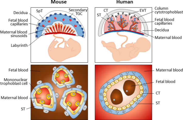
FIGURE 2.

Structure of human and mouse mature placenta. Structure of the mouse placenta. The inset details the fetal‐maternal interface in the labyrinth. Structure of the human placenta. The inset image shows a cross section of a chorionic villus; trophoblast‐derived structures (blue) and mesoderm‐derived tissues (orange). The inset images illustrate the number and type of cell layers between the maternal and fetal blood. CT, cytotrophoblast; EVT, extravillous cytotrophoblast; SpT, spongiotrophoblast; ST, syncytiotrophoblast; TGC, trophoblast giant cell
