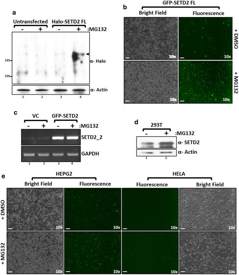
Fig. 1.
SETD2 is robustly degraded by the proteasome. a Western blot of whole-cell lysates probed with the depicted antibodies. Lysates of wild type 293T (untransfected) cells or expressing SETD2 full-length (FL) were prepared after 12 h of MG132 (10 µM) treatment. The expected band for the target protein is indicated by an arrow. b Microscopy images showing the effect of MG132 treatment on expression of GFP-SETD2 FL in 293T cells. The scale bar is 1 mm. c RNA was isolated from GFP-SETD2 FL transfected cells and RT-PCR was performed to check the transcript levels. GAPDH was used as a normalization control. VC- empty vector control. d Whole-cell lysates of 293T cells were prepared after 12 h of MG132 (10 µM) treatment and probed with the antibodies depicted. e Microscopy images showing the effect of MG132 treatment on the expression of GFP-SETD2 FL in the cell lines described. The scale bar is 1 mm

