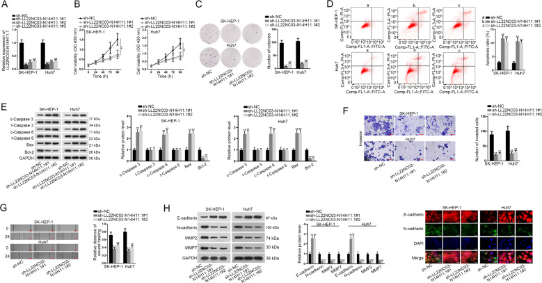
Fig. 2. Biological function of LL22NC03-N14H11.1 silence in HCC cells.
a RT-qPCR data of LL22NC03-N14H11.1 level in SK-HEP-1 and Huh7 cells transfected with sh-NC or sh-LL22NC03-N14H11.1#1/2/3. b Cell viability of SK-HEP-1 and Huh7 cells at indicated time was evaluated by CCK-8 under indicated transfection. c Representative images and quantification of colonies formed by two HCC cells under indicated transfection. d Flow cytometry analysis of apoptotic HCC cells under LL22NC03-N14H11.1 silence vs. control. e Western blots of cleaved and total caspase-3 and caspase-6, Bax, and Bcl-2 in 2 HCC cells. f Representative pictures of invading HCC cells and quantification under LL22NC03-N14H11.1 depletion were obtained via transwell assay. Scale bar: 100 μm. g Images of wound width of HCC cells of each group and quantification of relative distance of wound healing obtained from wound healing assay. h Western blots of E-cadherin, N-cadherin, MMP2, and MMP7 in HCC cells with LL22NC03-N14H11.1 silence. i IF staining of E-cadherin and N-cadherin in HCC cells of each group. Scale bar: 50 μm. **P < 0.01.

