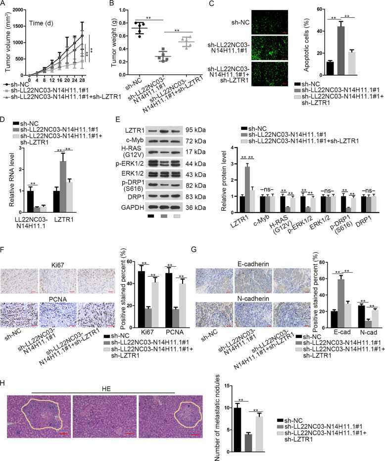
Fig. 7. LL22NC03-N14H11.1 drove tumorigenesis and metastasis in HCC through LZTR1 in vivo.
SK-HEP-1 cells were, respectively, transfected with sh-NC, sh-LL22NC03-N14H11.1#1, or sh-LL22NC03-N14H11.1#1 + sh-LZTR1 and injected subcutaneously or from tail vain to monitor tumorigenesis and metastasis of HCC. a Volumes of xenografts in mice of each group was evaluated every 4 days after subcutaneous injection and the growth curve was outlined. b Twenty-eight days after injection, tumors from each group were resected and weighed. c TUNEL staining was used to evaluate the apoptosis in tumors of each group. d RT-qPCR analysis of LL22NC03-N14H11.1 and LZTR1 in tumors of each group. e Western blot was implemented to detect the levels of LZTR1, c-Myb, H-RAS (G12V), p-ERK1/2, total ERK1/2, p-DRP1 (S616), and total DRP1 in tumors of each group. f, g IHC staining and quantification of Ki67, PCNA, N-cadherin, and E-cadherin in tumors of each group. h Metastatic nodules in tumors of each group was stained by HE and quantified. Scale bar: 100 μm. **P < 0.01; n.s. no significance.

