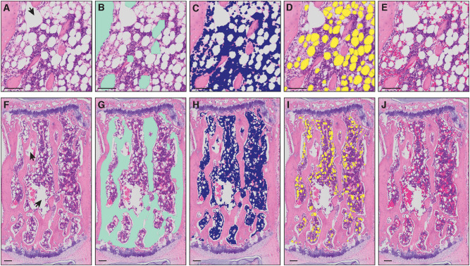
Figure 1.
Masks of bone marrow compartment areas detected by MarrowQuant. (A–E) Proximal tibia and (F–J) caudal vertebra (Cd2). Arrowheads in (A,F) represent manually excluded artefacts. (A,F) Unprocessed H&E image, (B,G) bone detection (green), (C,H) nucleated cell detection (violet), (D,I) adipocyte ghost detection (yellow), (E,J) interstitium and microvasculature (pink). Image from the proximal tibia at day 15 post lethal irradiation and total bone marrow transplant (A) and caudal vertebra 2 (F) of a C57BL/6 2-months-old female mouse housed at room temperature fed a standard ad libitum chow diet. Scale bars are 50 μm (A–E) and 100 μm (F–J). MarrowQuant user-defined parameters set at recommended values for bone marrow analysis (minimum adipocyte size: 120 μm2, maximum adipocyte size: 5,000 μm2, minimum circularity: 0.3, exclude edges: false).

