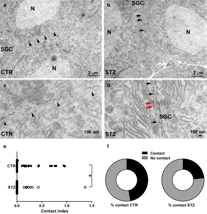
FIGURE 5.

Ultrastructural analysis of IB4+ DRG neurons in control and diabetic mice. (a) In CTR, the membranes of two adjacent clustered IB4+ sensory neurons are juxtaposed, without the interposition of glia (arrowheads). (b) In STZ‐treated mice, a glial sheath is present between two IB4+ neurons of the same cluster (arrows). (c) High magnification of (a). Note the occurrence of 20‐nm gold particles indicative of IB4 immunogold staining scattered over the entire length of the juxtaposed neuronal membranes (arrowheads). (d) High magnification of (b). Note the glia separating the membranes of two IB4+ DRG neurons (arrows) and a gap‐junction between the neuron and SGC (red double arrowheads). (e) Contact index in vehicle‐treated mice and STZ‐treated mice. The contact index is markedly reduced in STZ (Mann–Whitney test, p < .01). (f) Pie charts showing the proportion of neuronal membranes exhibiting at least one point of contact (Fisher exact test, p < .05) in CTR‐ and STZ‐treated mice. CTR, vehicle‐treated mice; N, nucleus; SGC, satellite glial cell; STZ, streptozotocin‐treated mice; *p < .05, **p < .01
