FIGURE 2.

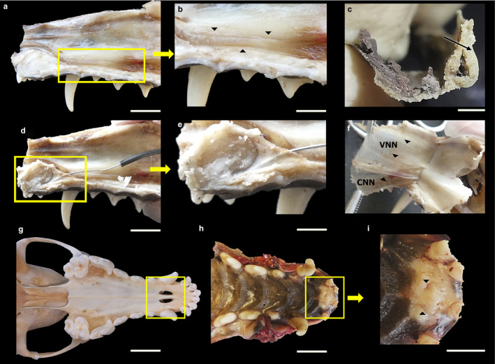
Dissection of the VNO and its innervation, showing the cannulation of the incisive duct and its opening in the incisive papilla, in the fox. (a) Lateral view of the nasal septum, where the VNO is framed. (b) Enlarged view of the nasal septum. The arrowheads delimit the VNO. (c) VNO extracted and cross‐sectioned, showing the lumen of the duct and the cartilaginous capsule (arrowhead). (d) Lateral view of the nasal septum. Cannulation of the incisive duct is framed. (e) Enlarged view of the cannulation. (f) Medial view of the nasal cavity mucosa after separation from the nasal septum. Innervation is indicated by arrowheads. VNN, vomeronasal nerves; CNN, caudal nasal nerve. (g) Ventral view of the skull of a fox. The palatine fissure is framed. (h) Ventral view of the hard palate. The rostral area is framed, and the higher magnification view shows that the incisive papilla can be found here, demarcated by arrowheads (i). Scale bars: (a,b,d,e,g,h) 1 cm, (c) 2 mm, (i) 0.5 cm.
