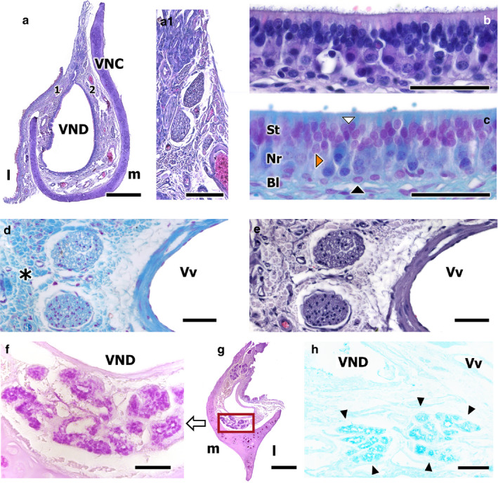
FIGURE 5.

Transverse sections of the fox VNO. (a) Haematoxylin‐Eosin (HE)‐stained transverse section of the VNO. Higher magnification views of the neuroreceptor epithelium, after HE (b), and Gallego’s Trichrome (c) staining, showing the three layers of the neuroepithelium, the sustentacular layer (St), the neuroreceptor layer (Nr), and the basal cell layer (Bl). The white arrowhead points to a sustentacular cell, the orange arrowhead to a neuroreceptor cell, and the black arrowheads to basal cells. Higher magnification of the unmyelinated nerves in (a.1) (HE‐stained). (d,e) Vomeronasal nerves. Gallego’s Trichrome and HE stains, respectively. Loose connective tissue is clearly identified (asterisk). (f–h) Disposition and morphology of the fox vomeronasal glands: ventral transversal section of the organ (f), which shows acinar, tubular, and acinotubular glands, serose and PAS+ in nature. PAS stain. VND, vomeronasal duct. (g) Transverse caudal section of the VNO, showing the PAS+ glandular tissue disposition. (h) Transverse caudodorsal section of the VNO. Tubular, serose, Alcian Blue+ glands. Alcian Blue stain. Scale bars: (a) 500 µm, (b–e) 50 µm, (f,h) 50 µm, (g) 250 µm
