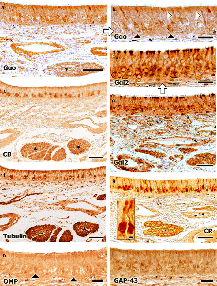
FIGURE 8.

Immunohistochemical study of the fox VNO neuroepithelium. (a) Immunopositive labelling with an anti‐Gαo antibody. A subpopulation of neurons is labelled, with labelling concentrated in the soma (black arrowheads, b) and in the dendrites (white arrowheads, b). A similar pattern is observed for the anti‐CB antibody (d). Widespread immunopositive labelling is observed for the anti‐Gαi2 antibody. All neuron components are strongly labelled: the soma, the terminal button, the axon, and the dendrites. The asterisks (d–g) indicate the vomeronasal nerve fascicles in the parenchyma (e, at higher magnification in c). This labelling pattern is similar to that observed for the anti‐CR antibody (g, an immunopositive neuroreceptor cell is framed). (f) Immunopositive labelling for the anti‐α‐tubulin antibody, showing an intermediate pattern between those observed for the anti‐Gαo and anti‐Gαi2 antibodies. (h,i) Immunopositive labelling for the anti‐OMP and anti‐GAP‐43 antibodies are shown, respectively. In comparison with the anti‐OMP antibody, broader labelling is observed for the anti‐GAP‐43 antibody, which suggests the regenerative ability of the neuroepithelium. Scale bars: (a,d–g) 50 µm, (b,c,h,i) 25 µm
