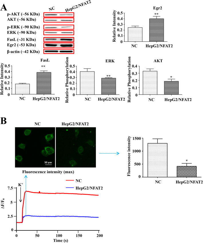
Fig. 3.
NFAT2 overexpression induced the expression of Egr2, FasL and suppressed the phosphorylation of AKT and ERK, and Ca2+ mobilization in HepG2 cells. a The protein expression of Egr2, FasL, AKT, p-AKT, ERK and p-ERK in NC and HepG2/NFAT2 cells, was detected by western blots (n = 3). b The top left pictures are representative fluorescence images in maximum relative fluorescence intensity of NC and HpeG2/NFAT2 cells and the top right bar graph is the statistical result of maximum fluorescence intensity in this two groups (n = 6). The bottom picture shows representative time-dependent relative fluorescence intensity changes of fluo-4-Ca2+ complex in NC and HpeG2/NFAT2 cells. Each bar represents the mean ± S.D.; *p < 0.05, **p < 0.01, ***p < 0.001 compared to the NC samples. Full-length blots are presented in Supplementary Figures

