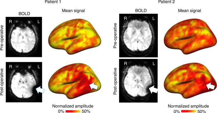
Fig. 7. Implanted electrode interference severely reduces BOLD signal amplitude.
Pre- and post-operative BOLD frames are shown in the left panels for two representative patients with Parkinson’s disease. Patients had electrodes implanted in the subthalamic nucleus, resulting in interference around left temporal, parietal, and occipital regions. The pre- and post-operative normalized and averaged BOLD signals are shown on inflated cortical surfaces in the right panels. Following surgery, the BOLD signal amplitude is severely affected in regions close to the electrode wires (temporal and parietal regions for patient 1; temporal, parietal, and occipital regions for patient 2).

