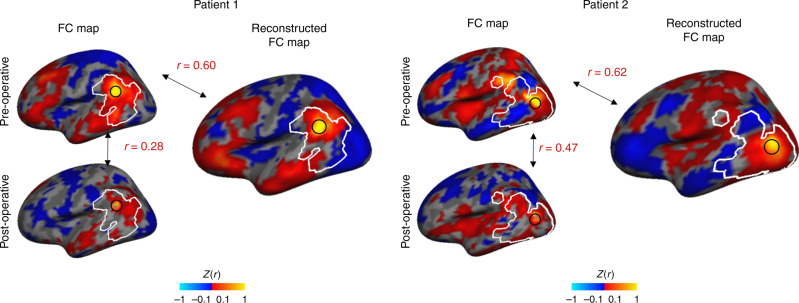
Fig. 8. The DCGAN model can reconstruct BOLD signals compromised by the implantation of a deep-brain stimulator in patients with Parkinson’s disease.
We generated the FC map of a given vertex (black circle) before and after surgery in two representative patients (left panels). Delineated in white is the region that is affected by the implanted device, defined by identifying the vertices which showed a stark decrease in BOLD amplitude after the implantation surgery. The post-operative FC maps are substantially different from the pre-operative FC maps due to the intracortical electrodes, with a relatively low correlation of r = 0.28 in patient 1 and r = 0.47 in patient 2. We also generated FC maps for the same vertices, this time using the reconstructed BOLD frames (right panels). The generated FC maps are highly similar to the intact pre-operative FC maps, as indicated by a correlation of r = 0.60 for patient 1 and r = 0.62 for patient 2. Thus, the DCGAN model is able to reconstruct patients’ functional brain organizations.

