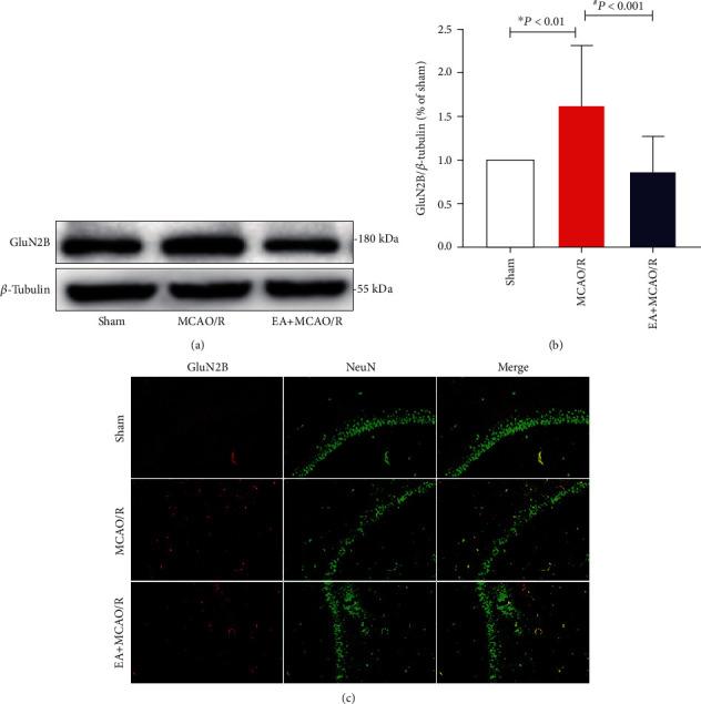
Figure 3.

Effect of EA pretreatment on GluN2B expression (n = 4). GluN2B protein expression and cell localization were detected at 24 h after cerebral I/R by WB and double immunofluorescence staining. (a) Representative WB bands showing GluN2B expression in the rat hippocampus of three groups. (b) Comparison of the GluN2B expression. The results conformed to the normal distribution with heterogenous variance. Differences among groups were examined using the Kruskal-Wallis test (H = 18.598, P < 0.001) followed by the Dunn test for post hoc multiple comparisons. (c) Representative double immunofluorescence staining (yellow) of GluN2B-positive cells (red) and NeuN-positive cells (green) in brain sections. Scale bars = 100 μm. ∗P vs. sham; #P vs. MCAO/R.
