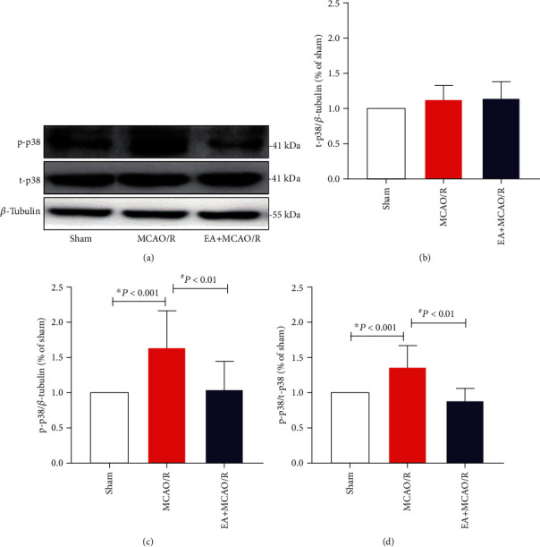
Figure 5.

Effect of EA pretreatment on t-p38 and p-p38 expression in the hippocampal CA1 neurons (n = 4). The expression of p38 and p-p38 in the hippocampal neurons was detected by WB at 24 h after cerebral I/R. (a) Representative WB bands showing p38 MAPK expression in the rat hippocampus of three groups. (b, c) Comparison of total p38 (t-p38) and phophorylated p38 (p-p38) expression. The result of t-p38/β-tubulin conformed to normal distribution with homogeneous variance. Differences among groups were examined using AVONA (F(2, 33) = 1.941, P = 0.160 > 0.05). The result of p-p38/β-tubulin conformed to normal distribution with homogeneous variance. Differences among groups were examined using AVONA (F(2, 33) = 10.380, P < 0.001). (d) Comparison of p-p38/t-p38 ratio. The result of p-p38/t-p38 conformed to normal distribution with homogeneous variance. Differences among groups were examined using AVONA (F(2, 33) = 18.015, P < 0.001) followed by LSD for post hoc multiple comparisons. ∗P vs. sham; #P vs. MCAO/R.
