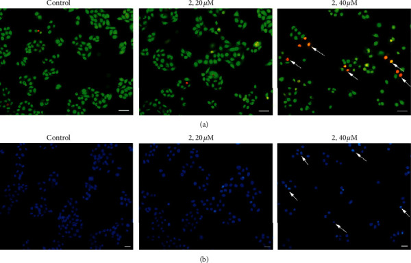
Figure 3.

Eca-109 cells were stained with AO/EB (a) and Hoechst 33342 (b) after being exposed to complex 2 (0, 20, and 40 μM) for 24 h under a fluorescence microscope. Representative photomicrographs from three independent experiments. Arrows indicate apoptotic bodies. Scale bars: 20 µm.
