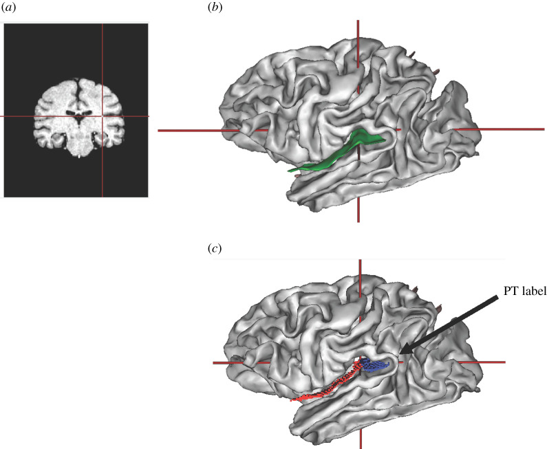
Figure 2.
(a) Coronal view of T1 can and (b) lateral view of the three-dimensional brain with the Sylvian fissure outlined in green. Note that the cross bars in each image reflect the location of the point of closure of the inferior limb of the insula, which served as the anterior border to define the PT. (c) Lateral view of the three-dimensional brain showing the division of the Sylvian fissure into the anterior (red) and posterior (blue) regions after using the scissors to bifurcate the fold. (Online version in colour.)

