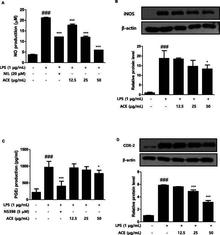
Fig. 1.
Effects of ACE on the levels of inflammatory mediators in RAW 264.7 macrophages. Cells were treated with 12.5, 25, or 50 μg/mL of ACE for 1 h prior to the addition of LPS (1 μg/mL), and the cells were incubated for 24 h and 48 h, respectively. a NO and (c) PGE2 level were figured out with Griess reagent and the EIA kit, respectively. NIL (20 μM) or NS398 (5 μM) was used as positive control. The protein level of (b) iNOS and (d) COX-2 were determined by western blot analysis with specific antibodies. Densitometric analysis was performed with Image J software (version 1.50i). Values are presented as mean ± S.D. of three independent experiments. ###p < 0.001 when compared with control; *p < 0.05, ***p < 0.001 when compared with LPS-induced group. Significant differences among treated groups were determined by ANOVA and Dunnett’s post hoc test

