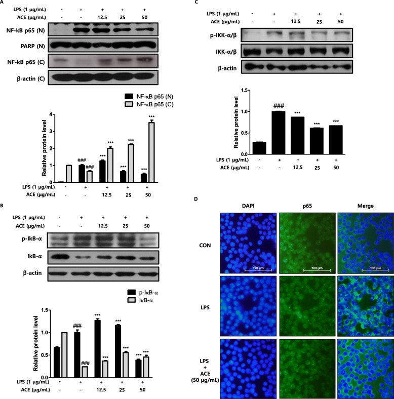
Fig. 3.
Effects of ACE on LPS-induced NFκB pathway in RAW 264.7 macrophages. a Cells were treated with 12.5, 25, or 50 μg/mL of ACE for 1 h prior to the addition of LPS (1 μg/mL). LPS stimulation took time on NF-κB p65 for 30 min. Nuclear (N) and cytosolic (C) extracts were isolated and adjusted for the detection of p65 with specific antibodies. After incubation with LPS for 15 min (b) and 5 min (c), total proteins were prepared and western blot assay was performed with specific antibodies. PARP and β-actin were shown as internal controls. Densitometric analysis was performed with Image J software (version 1.50i). Data are presented as mean ± S.D. of three independent experiments. ###p < 0.001 when compared with control; ***p < 0.001 when compared with LPS-induced group. Significant differences between treated groups were determined by ANOVA and Dunnett’s post hoc test. d Cells were pre-treated with ACE for 1 h prior to the addition of LPS for 30 min. The nuclear translocation of NF-κB p65 was visualized by immunofluorescence. The nuclei were counterstained with DAPI (blue). The stained cells were visualized with a fluorescence microscope at 400X magnification

