FIGURE 5:
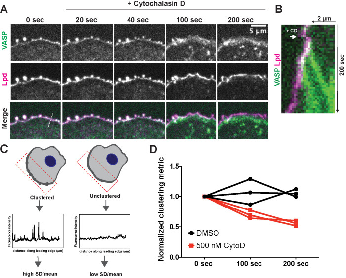
Free barbed ends of actin filaments are required for VASP/Lpd cluster stability. (A) Representative example of a B16F1 cell expressing VASP-eYFP (green) and Lpd-tdTomato (magenta) before and after acute treatment with 500 nM cytochalasin D. (B) Kymograph of a slice taken perpendicular to the leading edge shows change in membrane localization of VASP (green) and Lpd (magenta) upon addition of cytochalasin D. (C) Schematic of quantification of changes in clustering. The SD along a defined leading edge before (0 s) and after drug treatment (100 s, 200 s) was calculated and normalized to the mean fluorescence intensity. (D) Quantification of Lpd clustering at foci following acute treatment with cytochalasin D or DMSO (vehicle) shows that free barbed ends are required for robust clustering of VASP and Lpd. Clustering is represented by SD along the leading edge ROI before and after treatment. Symbols are averages from three biological replicates, each with >8 cells. Paired t test of unnormalized data: p = 0.02.

