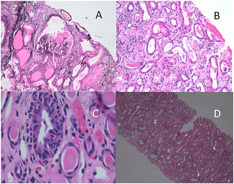
Figure 1.
(A) Periodic acid–Schiff (PAS) stain showing epithelial proliferation and compression of the glomerulus. (B) Hematoxylin and eosin (H&E)-stained section showing dilated tubules with flattened epithelial cells; and cells and debris within tubular lumena. (C) H&E-stained section showing intraepithelial neutrophils. (D) H&E-stained section photographed with polarized light showing crystalline material within tubules.

