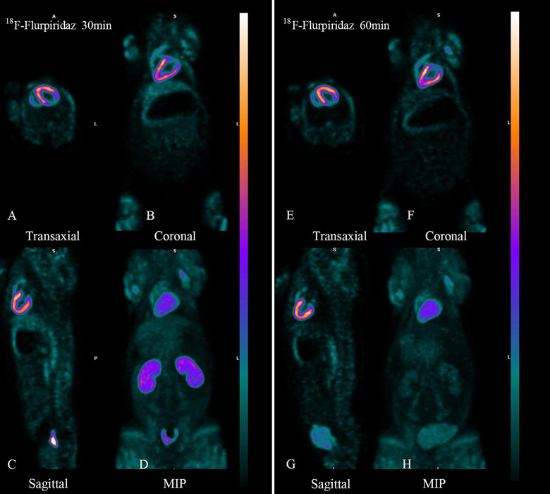
Figure 2.
Body imaging of 18F-Flurpiridaz 30 min (left) and 60 min (right) in normal miniature pigs. The left and right of the images were 30min and 60min body images with 18F-Flurpiridaz PET/CT, respectively. A and E are transverse slices, B and F are coronal slices, C and G are sagittal slices, and D and H were maximum intensity projections(MIPs). No matter whether 30min or 60 minutes, left ventricular myocardium was clearly delineated and there was no radioactive interference outside the heart, and the radioactivity in cardiac cavity, lung tissue and liver was very low, and the radioactivity in kidney was significantly decreased in 60min.

