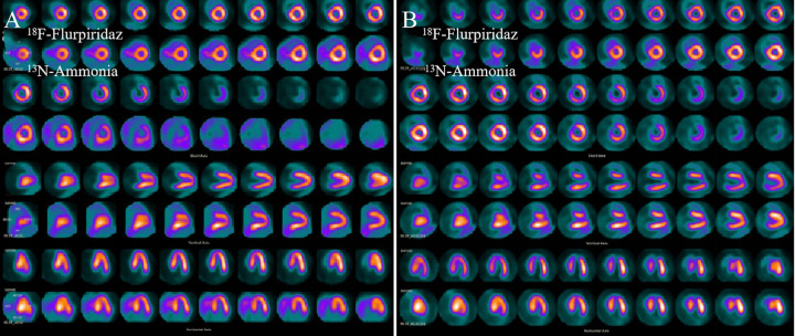
Figure 5.
Comparison of MPI between 18F-Flurpiridaz and 13N-NH3·H2O in normal and infarct miniature pigs. From top to bottom, there were short axis (1 to 4 rows), vertical long axis (5 to 6 rows) and horizontal long axis (7 to 8 rows). The odd row was 18F-Flurpiridaz images and the even row was 13N-NH3·H2O images. a (normal group): the myocardium of each wall of the left ventricle was clearly delineated. Compared with 13N-NH3·H2O imaging, the 18F-Flurpiridaz distribution in the myocardium was very uniform and the myocardial walls were well displayed. b (infartion group): 18F-Flurpiridaz clearly delineated apical and part of anterior wall apical infarct area (radioactive defect), myocardium and infarction boundary were clearly delineated, 13N-NH3·H2O image showed a small amount of radioactivity distribution outside the heart.

