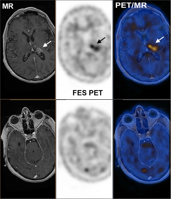
Figure 2.

MR (left), 18F‐FES PET (center), and fused PET/MR (right) of a patient with brain metastases from a historically ER‐positive breast cancer. The upper panel illustrates a lesion evident on MR, whereas the lower panel illustrates an 18F‐FES‐positive lesion that was not readily apparent on MR.
Abbreviations: 18F‐FES, 16α‐18F‐fluoro‐17β‐estradiol; MR, magnetic resonance; PET, postitron emission tomography.
