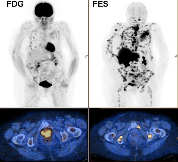
Figure 3.

Transaxial pelvic (at the level of the pubic symphyses) positron emission tomography (PET)/computed tomography fusion images (18F‐FDG [left] and 18F‐FES [right]). The insets represent the whole‐body PET image, of a patient with metastatic lobular ductal carcinoma of the breast. Note that 18F‐FDG uptake in many lesions is less than corresponding 18F‐FES uptake.
Abbreviations: 18F‐FDG, 18F‐fluorodeoxyglucose; 18F‐FES, 16α‐18F‐fluoro‐17β‐estradiol.
