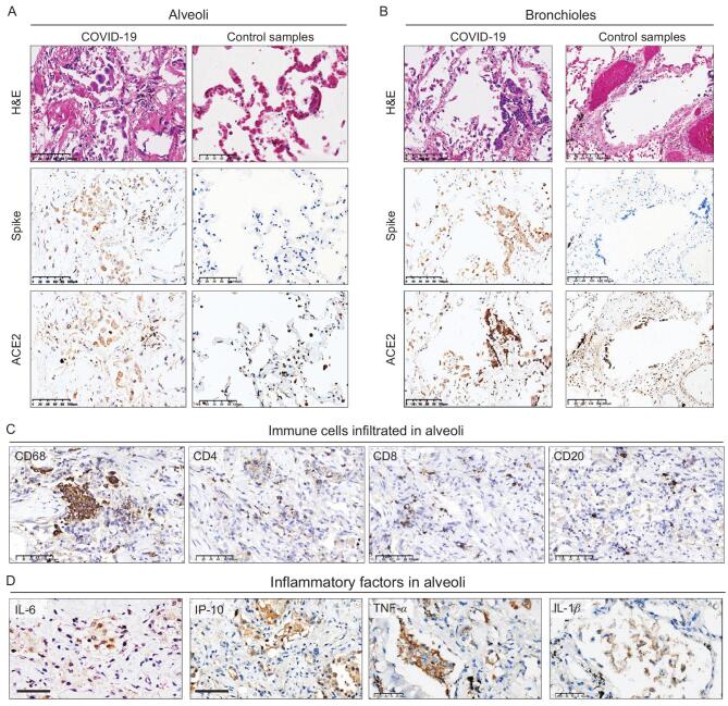
Figure 2.
Pulmonary immune signature in COVID-19 autopsy patients. (A and B) IHC staining of SARS-CoV-2 spike protein, ACE2, and the corresponding H&E staining from the same microscopic field using serial sections in alveoli (A, Case 1) and bronchioles (B, Case 2) from COVID-19 cases and control lung tissues from a non-COVID-19 case who died from heart attack (C) IHC staining of macrophages marked by CD68 and lymphocytes marked by CD4, CD8, CD20 in alveoli (Case 3). (D) IHC staining of inflammatory factors (IL-6, IP-10, TNF-α, IL-1β) in serial sections of alveoli (Case 6). Scale bar = 100 μm (A, left panels of B and C), 200 μm (right panels of B) or 50 μm (D).

