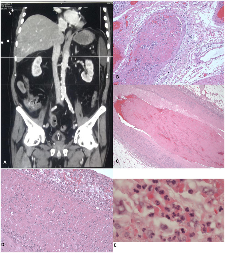
Fig. 1.
Arterial and venous thrombotic complications and neutrophilic vasculitis in splenic artery
(A) Thrombosis of the coeliac tripod immediately after its origin, extended to ∼15 mm. The superior mesenteric artery presents thrombosis as well as some of its branches. Almost complete infarction of an ileal segment and spleen. (B–E) All anatomic specimens were fixed in 4% neutral buffered formaldehyde and, after paraffin embedding, 3 micra thick sections were cut and routinely stained with haematoxylin and eosin (HE). (B) Venous vessel of the splenic hilum with fibrin thrombus in the lumen. HE, ×100 (original magnification). (C) Wall of the splenic artery with fibrin thrombus in the lumen and granulocytic infiltration. HE, ×100 (original magnification). (D) High magnification of the splenic artery showing transmural infiltration of neutrophils, from adventitia (top) to intima (bottom left). HE, ×200 (original magnification). (E) Diffuse infiltration of neutrophilic granulocytes in the arterial wall. HE, ×1000 (original magnification).

