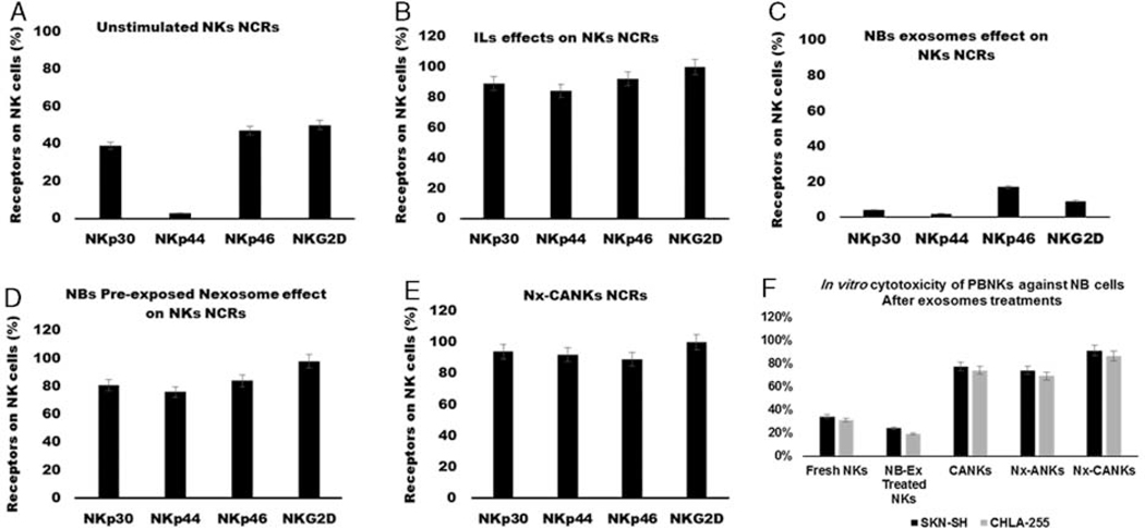
FIGURE 5.
Natural cytotoxicity receptors on NK cells. A, NK cells were stained with specific NCRs monoclonal antibodies, a resting NK cell expresses different levels of NCRs. Less than 40% of NK cells express NKp30, and the level of NKp44 is undetectable. B, Cytokine-activated NK cells express high levels of NCRs. C, NB-Ex reduces the levels of all NCRs under the resting state of NK cells. D, The Nx-ANKs induce the expression of NCRs especially NKp44 similar to CANKs. E, Nexosomes have a synergistic effect on CANKs and induce the maximum expression of all NCRs that prepare NK to have a cytotoxic function against neoplastic cells. F, In vitro cytotoxicity of peripheral blood natural killer cells against NB cells. Nexosomes strongly stimulated NK activity in the presence of IL-21. IL indicates interleukin; NB, neuroblastoma; NB-Ex, NB-derived exsosomes; NK cell, natural killer cells; Nx-ANKs, NB exosome–activated NK; Nx-CANKs, NB exosome cytokine-activated NKs.

