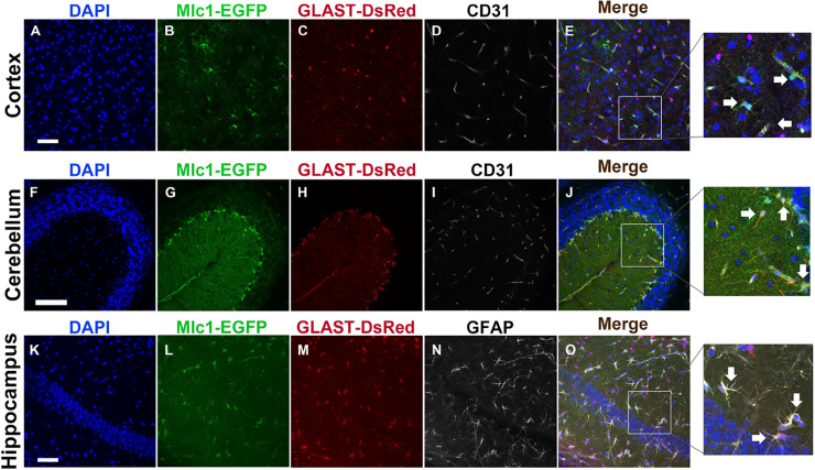
Fig 3. Analyzing PA and non-PA cell distribution in adult Mlc1-EGFP;GLAST-DsRed mice based on EGFP and DsRed reporter expression.
(A-E); Coronal brain sections through the cerebral cortices of an adult Mlc1-EGFP; GLAST-DsRed mouse brain were labeled with DAPI (A), anti-GFP to detect PAs (B), anti-RFP to detect DsRed-expressing PAs and non-PAs (C), and anti-CD31 to detect vascular ECs that comprise blood vessels (D). Note that cells adjacent to cortical blood vessels (PAs) co-express GFP and DsRed (arrows in E), whereas DsRed+ cells (non-PAs) that are more distal to CD31+ ECs lack GFP expression. Scale bars, 100 μm (A) and 20 μm (inset, E). (F-J); Coronal sections through the cerebellum of an adult Mlc1-EGFP;GLAST-DsRed mouse brain stained with DAPI (F), anti-GFP (G), anti-RFP (H), and anti-CD31 (I). Note that most if not all cerebellar Bergmann glial cells, which are a radial glial-like astrocyte sub-population, co-express GFP and DsRed (arrows in J). Scale bars, 100 μm (E) and 20 μm (inset, J). (K-O); Coronal sections through the hippocampus of an adult Mlc1-EGFP;GLAST-DsRed mouse brain labeled with DAPI (K), anti-GFP (L), anti-RFP (M), and anti-GFAP (N). Note that many hippocampal astrocytes co-express GFP, DsRed, and GFAP (arrows in O). Scale bars, 100 μm (K) and 20 μm (inset, O).

