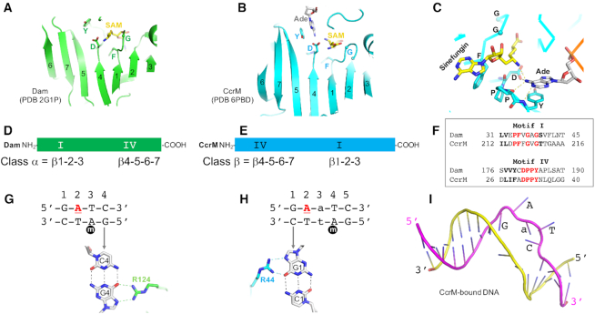
Figure 2.
Comparison of Dam (class α) and CcrM (class β). (A, B) The catalytic domain of the seven-stranded β structure in Dam (panel A) and CcrM (panel B). (C) Interactions with the SAM–adenosyl and DNA–adenosyl moieties in the active-site of CcrM. The phenylalanine of motif I provides an edge-to-face interaction to the face of SAM-adenosyl ring. The DPPY motif interacts with DNA adenine. (D–F) The locations of motif I and motif IV are reversed in the amino acid sequences of Dam and CcrM. (G) Dam interacts with guanine G4 of the non-target strand. (H) CcrM interacts with guanine G1 of the target strand. The underlined letter A in red is the methylation target. (I) Two strand separation in the CcrM-bound DNA.

