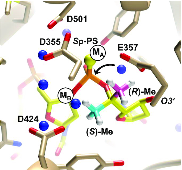
Figure 11.

Crystal structure of the KF exo active site bound to 3′-d(TPSTTT)-5′, where PS indicates an Sp phosphorothioate linkage in which 5′-(R)-C-methyl (pink) and 5′-(S)-C-methyl (cyan) groups were inserted on the 3′-terminal nucleotide. Positions of Zn2+ ions in the crystal structure of KF exo with native DNA but absent in the structure with a Sp-PS-modified oligo are indicated by black circles labeled MA and MB, water molecules are blue spheres, and an arrow indicates attack by a water molecule at the 3′-terminal phosphate group.
