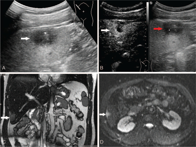
Figure 1.

A: A hypo echoic lesion with a size of 2.5 cm x 1.8 cm located in the sixth segment of liver was detected on the transabdominal US. B: The lesion showed hyper enhancement on early arterial phase (white arrow) and quickly wash-out on late arterial phase (red arrow) after intravenous injection of sulfur hexafluoride-filled micro bubble contrast agent. C: A hyper intensity lesion with a size of 2.1 cm x 1.7 cm on T2-weight image of MRI was presented. D: The hyper intensity lesion was partly diffusion restricted on diffusion-weighted MR images.
