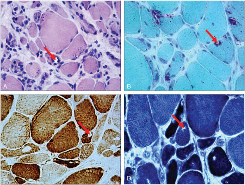
Figure 2.

Histopathological examination of the skeletal muscles. (A) Hematoxylin and eosin staining shows muscle fibers of variable sizes and rimmed vacuoles (red arrow). (B) Modified Gomori trichrome staining shows rimmed vacuoles in the muscle fibers (red arrow). Staining for (C) cytochrome oxidase and (D) NADH-tetrazolium reductase shows some fibers with areas lacking enzyme reactivity. (red arrow). (magnification, × 200).
