
Figure 1.
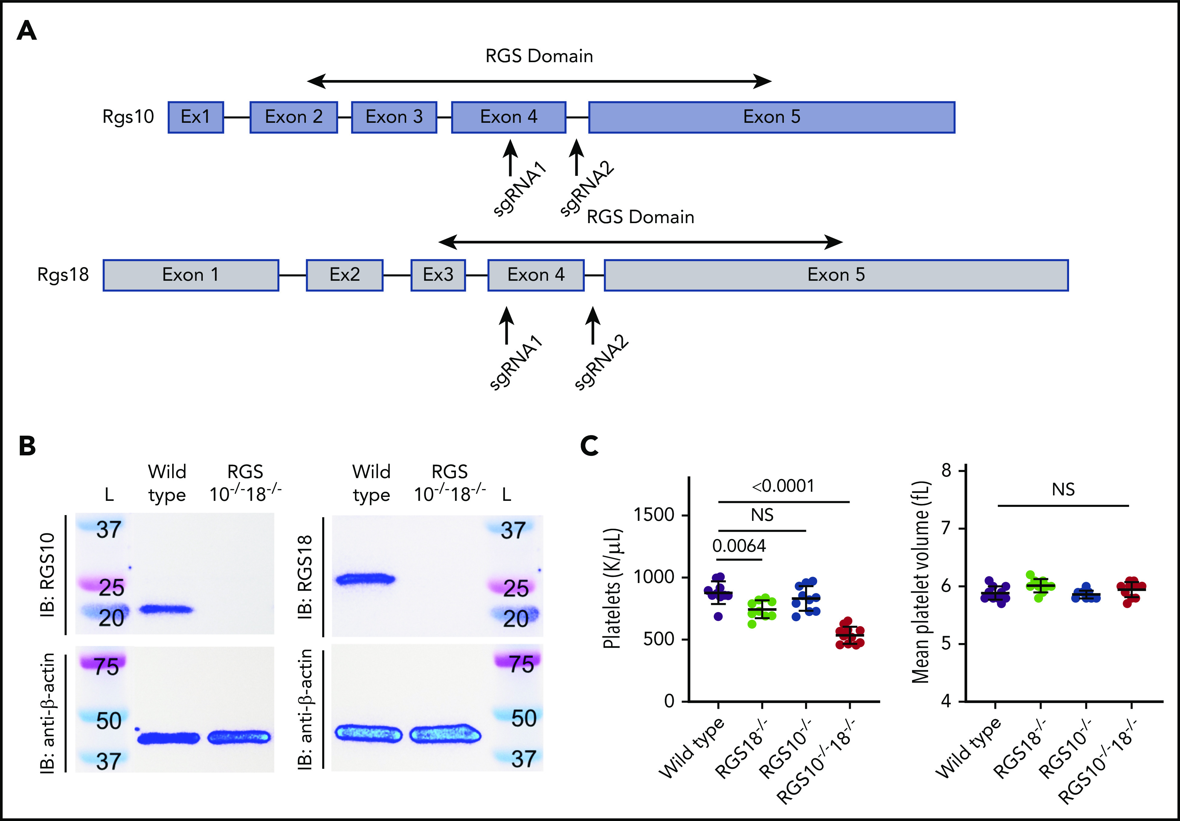
Generation and characterization of Rgs deletion mice. (A) Graphical depiction of Rgs10 and Rgs18 genes. Arrows indicate approximate locations targeted by single guide RNAs during CRISPR-Cas9. In both cases, regions within the sequence that encode the RGS domain were targeted. (B) Representative RGS10 and RGS18 immunoblots (IB) (top) of platelet lysates from RGS10+/+18+/+ (denoted “Wild type”) and RGS10−/−18−/− mice with β-actin (bottom) as the loading control. (C) Platelet counts and mean platelet volume of 8-week-old WT, RGS18−/−, RGS10−/−, and RGS10−/−18−/− mice. At least 9 measurements were collected per genotype. NS indicates P > .05; mean ± SEM.