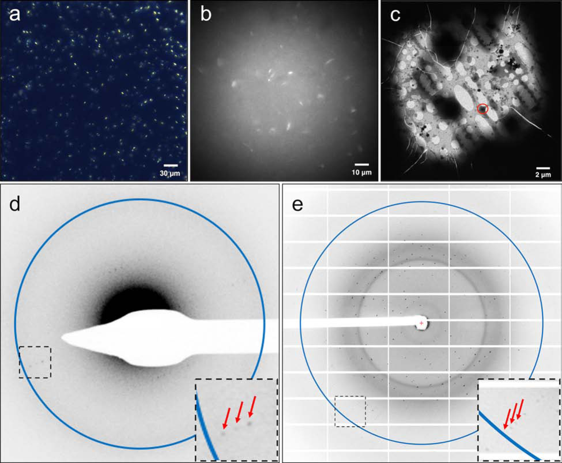
Figure 5. A2AAR microcrystals monitoring and MicroED diffraction.

A2AAR were crystallized in LCP to a size of 5x5x2 μm3 (a, viewed with cross-polarized light), and microcrystals were survived after LCP phase conversion by the converting buffer supplemented with 7% MPD (b, viewed with UV). Microcrystals were located on the EM grids (c) and a still initial electron diffraction pattern with resolution ring of 4.5 Å (d) was recorded before the continuous-rotation MicroED data collection in cryo-TEM. Red arrows denote the diffracted spots to 4.5 Å in a closer view of the black boxed area. (e) A2AAR microcrystals, treated with the same phase conversion method, retained their diffraction power to ~2.4 Å resolution (shown as the resolution ring in the image) at a microfocus X-ray beamline (diffracted spots to the highest resolution were denoted by red arrows in a closer view of the black boxed area).
