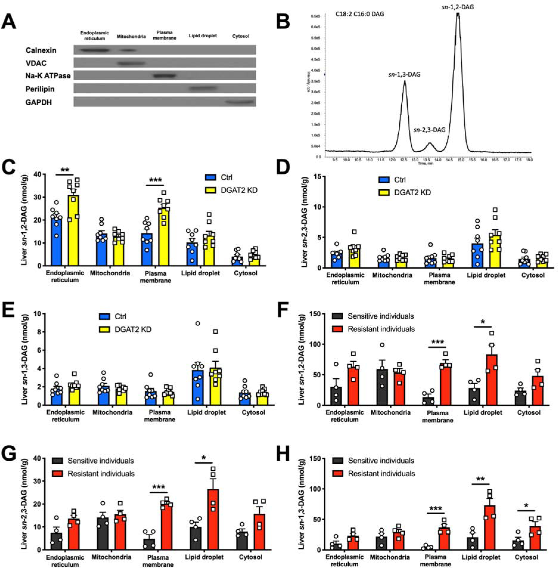
Figure 2. Liver PM sn-1,2-DAG Content Tracks with HIR in Rats and Humans.

(A) Separation of five subcellular compartments in liver measured by western blot.
(B) Representative chromatogram (n = 13) of DAG stereoisomer separation on LC/MS-MS.
(C), (D) and (E) Liver DAG stereoisomer content in five subcellular compartments in Ctrl vs DGAT2 KD rats.
(F), (G) and (H) Liver DAG stereoisomer content in five subcellular compartments in human individuals who were insulin-sensitive (black) or insulin-resistant (red).
In all panels, data are the mean±S.E.M. In (C), (D) and (E), n = 8 per group. In (F), (G) and (H), n = 4 per group. *P < 0.05, **P < 0.01 and ***P < 0.001.
