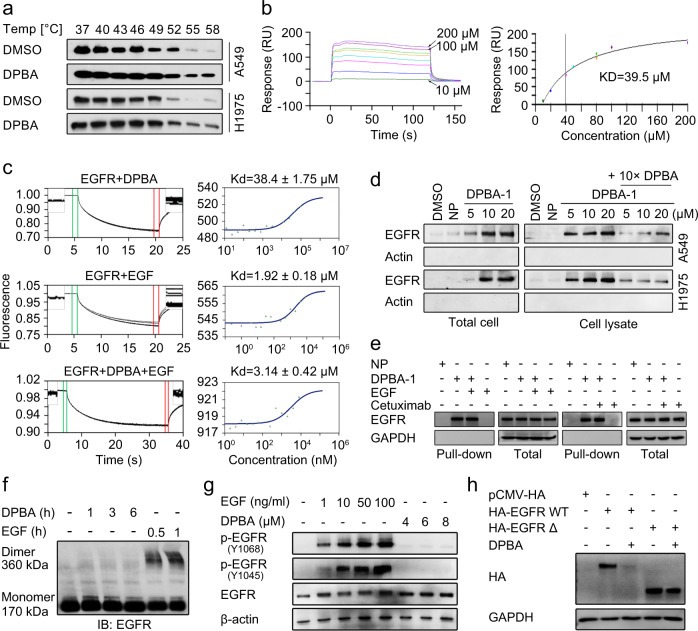
Fig. 4.
DPBA directly binds to EGFR ECD. a DPBA thermally stabilized EGFR in cellular level. A549 and H1975 were treated with DPBA (6 μM) for 3 h. Lysates were divided into eight fractions, followed by heating to indicated temperatures. Soluble EGFR was detected by Western blot. Interaction between DPBA (200, 100, 80, 80, 50, 40, 20, and 10 μM) and EGFR ECD was measured by BIAcore (b) or MST (c). d DPBA-1 dose-dependently interacted with EGFR in both total cell and cell lysate. In the total cell assay, A549 and H1975 were treated with NP (20 μM) or indicated concentrations of DPBA-1 for 1 h. In the cell lysate assay, A549 and H1975 were lysed, followed by indicated treatment. Interactions between EGFR and DPBA-1 were detected by pull-down assay. e EGF or cetuximab did not block the interaction of EGFR and DPBA-1. A549 was treated with DPBA-1 (10 μM) in the presence or absence of EGF (10 ng/ml) or cetuximab (5 μg/ml) for 1 h. Interactions between EGFR and DPBA-1 were detected by pull-down assay. f DPBA did not induce EGFR dimerization. A549 was treated with DPBA (6 μM) for 1 h, 3 h, and 6 h, or with EGF (200 ng/ml) for 0.5 and 1 h. EGFR dimerization was detected by Western blot. g A549 was treated with indicated concentrations of EGF or DPBA for 30 min. Total EGFR, p-EGFR (Y1068), and p-EGFR (Y1045) were measured by Western blot. h Deletion of EGFR ECD impaired DPBA-induced EGFR degradation. HEK-293T transfected with pCMV-HA, HA EGFR WT, or HA EGFR Δ was treated with DPBA (6 μM) for 24 h, and HA expression level was measured by Western blot

